Sawchyn Medical Illustration

Key Details
-
Project Date: 22.12.2012
-
Project Budget: 10.000 USD
-
Technologies: PHP, HTML, CSS, JS
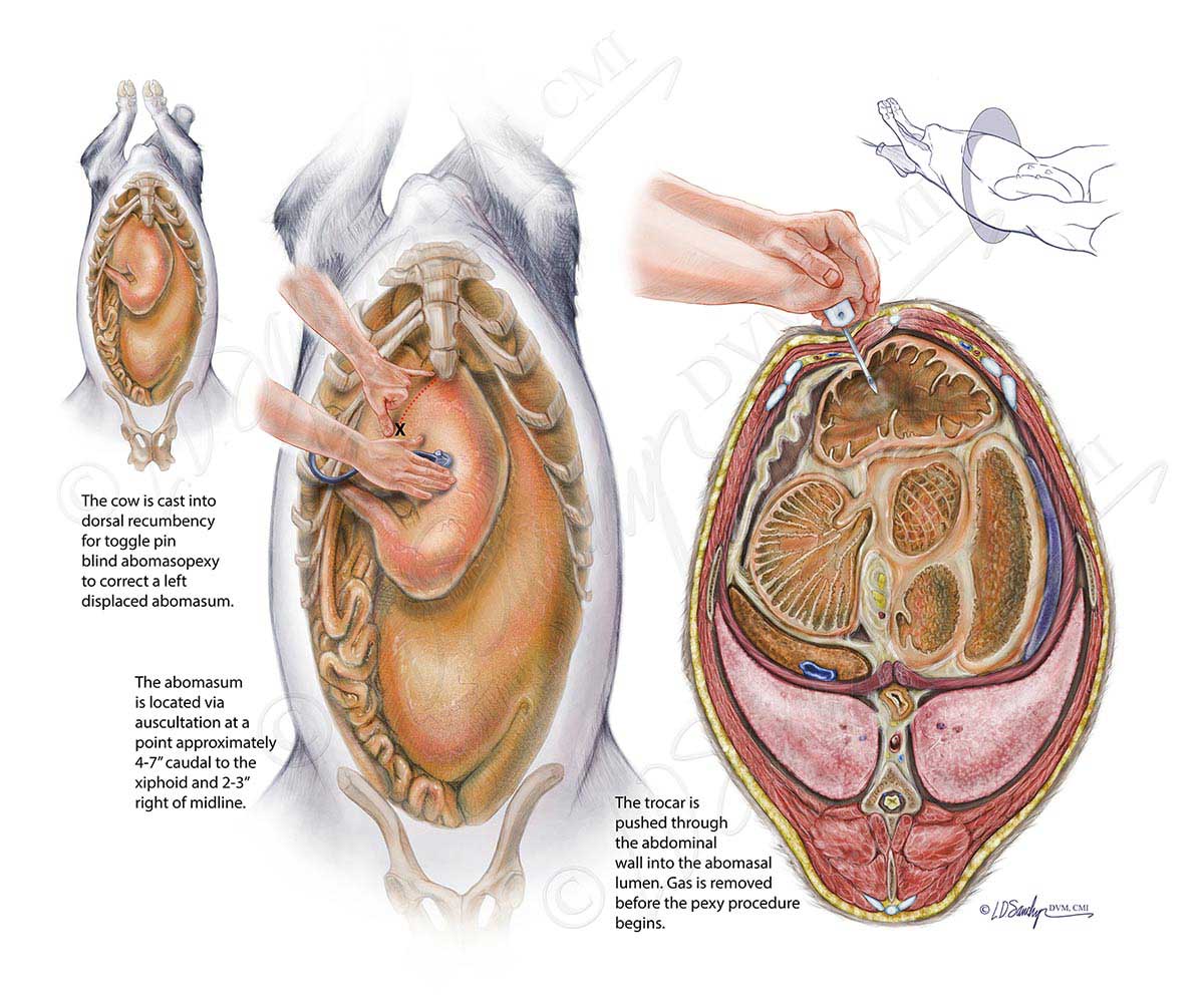
These color didactic illustrations were created to assist veterinarians in visualization of anatomy prior to placement of toggle pins to correct bovine left displacement of the abomasum (LDA). Photographs and videos of the procedure were inadequate to show the complex anatomy, and using a cadaver for the procedure was not ideal. During LDA, the abomasum relocates from its normal position, right of the ventral midline, to between the rumen and left body wall (not shown). Toggle pin stabilization, which involves casting the animal into dorsal recumbency and suturing the abomasum to the ventrum, is a minimally invasive repair in a stable patient.
In dorsal recumbency, trapped gas causes the abomasum to shift back to a “normal” position, located by simultaneous auscultation and percussion of the ventral abdomen to detect a tympanic “ping”. After placement of the trocar (shown), toggle pins attached to suture are placed into the abomasal lumen through the trocar. Tying of the sutures and subsequent scar formation holds the abomasum in place.
We Accept PayPal
![]()